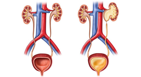
Hydronephrosis
Hydronephrosis is a condition in which urinary retention occurs in the renal collecting system, and is reflected in its enlargement. The renal pelvis (pylon) and the renal calyces are enlarged. In the case of major enlargement, pressure on the kidney tissue (parenchyma) occurs. The most common cause of hydronephrosis is a narrowing (stenosis) of the pyeloureteral junction (a joint of the renal pelvis and the ureter).
What is stenosis of the pyeloureteral junction?
The stenosis of the pyeloureteral junction is a narrowing of the junction of the renal pelvis (pyelone) and the ureter. The most common reason for this is an obstruction in the normal flow of urine from the kidney to the ureter. When it comes to the congenital conditions, it can usually be seen prenatally, during an ultrasound examination. It can also result from infection or trauma.
Stenosis of the pyeloureteral (PU) compound can also be spontaneously repaired. If not diagnosed during pregnancy, infections of the kidney system - pyelonephritis - may occur in early childhood. Stenosis of the PU junction over time leads to dilatation of the pyelocalyx system and the formation of hydronephrosis - accumulation of urine in the canal system of the kidney due to the obstruction of urine output. Hydronephrosis can lead to loss of a kidney function.
What are the symptoms of stenosis of the pyeloureteral compound?
The stenosis of a PU compound may not necessarily have symptoms and sometimes manifest itself for the first time only in advanced school age. Acute abdominal pain or chronic inflammation of the lower back and abdominal muscles can be seen if stenosis "triggers". Such pain may resemble renal colic and occur after excessive fluid intake. Pain can occur during urination, and there may be nausea and vomiting. Patients see their doctor for frequent urinary infections (pyelonephritis) or when they see blood in their urine (hematuria).
How is PU stenosis diagnosed?
The first step in making a diagnosis is a history. Some of the diagnostic tests and tests are:
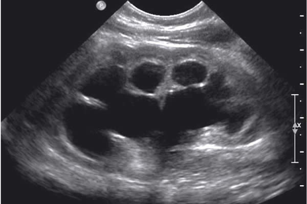
- Abdominal ultrasound and ultrasound
- Magnetic urography
- Kidney scintigraphy
- Micturating cystourethrography is performed to exclude urine reflux (return) to the kidney
How is this condition treated?
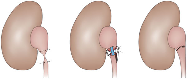
Treatment of this condition requires corrective surgery called pyeloplasty. The purpose of the treatment is to improve urinary flow and prevent further damage to the kidney parenchyma. Sometimes it is necessary to place a stent, catheter, or percutaneous nephrostomy immediately in order to at least temporarily eliminate the symptoms of urinary blockage.
The goal of pyeloplasty is to remove the narrowed part of the ureter. The operation is performed under general anesthesia. At the end of the procedure, an internal ureteral stent, also known as a double J stent, is placed. After one month of surgery, the stent is removed and treatment is terminated.
Complications may be related to anesthesia or specific complications. Possible occurrence of bleeding, infection, occurrence of postoperative hernia in the incision area, damage to surrounding organs, leakage of urine on the drain that is placed during surgery.
The return to normal physical activities is four weeks after surgery.